OS TRIGONUM SYNDROME
OS TRIGONUM SYNDROME
INTRODUCTION:
v Up to 40 accessory ossicles
and multiple sesamoids have been described in the foot and ankle
· Definition
o
accessory
ossicles are secondary ossification centers that
remain separated from the normal bone
o
sesamoids
are bones that are incorporated into tendons and move with
normal and abnormal tendon motion
v Most common ossicles
· os trigonum
· accessory navicular (os
tibiale externum)
· os intermetatarseum
v Most common sesamoids
· os peroneum
· located in the peroneus
longus tendon
· hallux sesamoids located in
the flexor hallucis brevis tendon at the base of the 1st metatarsal head.
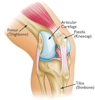
ANATOMY:

DEFINITION:
Accessory ossicle representing
the separated posterolateral tubercle of the talus usually asymptomatic, but
can become symptomatic and cause os trigonum syndrome.
EPIDEMOLOGY:
Incidence 10-25% of the
population have os trigonum commonly symptomatic
in ballet dancers due to extreme plantar flexion ("en
pointe" toe position)

PATHOPHYSIOLOGY:
Pathophysiology
of os trigonum syndrome repetitive microtrauma (ankle plantarflexion) may present as a stress fracture acute forced
plantarflexion may present as an acute fracture.
OSTEOLOGY:
The secondary ossification
center forms posterior to the talus between 8-13yrs normally fuses with talus within 1yr if the ossicle fails to
fuse, it articulates with the talus through a synchondrosis the os lies lateral to FHL, tibial
nerve, PTT, and posterior tibial artery.
SYMPTOMS:
Symptoms pain in "en pointe"
position physical exam posterolateral ankle pain with
passive ankle plantar flexion differentiate from FHL
tendinitis where ankle pain is posteromedial and there may be
triggering may have swelling and tenderness over FHL if
associated with FHL tendinitis.
IMAGING:
X-Ray:

MRI:

TREATMENT:
§ Non-operative:
§ NSAIDS,
rest, immobilization, restricted weightbearing
§ Operative:
§ Surgical
excision:
§ Indications: if non-operative management fails
Techniques:
through open lateral approach or posterior ankle arthroscopy.


Comments
Post a Comment
Thank you for your kind words and your support.