Coronoid Process Fracture - Case Presentation
CASE PRESENTATION
INTRODUCTION & BACKGROUND:
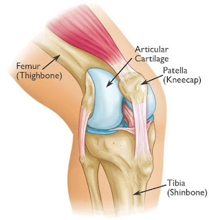
The coronoid is the most important portion of
ulno-humeral articulation
Reasons:
1. Provides anterior
buttress
2. Anterior capsule and
brachialis attach to coronoid
3. Anterior band of the
MCL attaches to it
- Distally and medially on sublime tubercle
Instability rises and prognosis deteriorates according to the amount of
coronoid process that is fractured.
ANATOMY:


CLASSIFICATION:
Coronoid
process fractures have been classified into three types:
·
type 1: avulsion
of the tip of the coronoid process
·
type 2: fragment
involving <50% of the coronoid process
·
type 3: fragment
involving >50% of the coronoid process
The prognostic
relevance of this classification is contentious, but there is some correlation
with the pattern of associated injuries: smaller fractures are more likely to
be associated with the “terrible
triad” pattern of injury, whereas larger fragments tend to occur
with anterior and posterior fracture-dislocations of the olecranon
PATIENT INFORMATION:
Ø Patient
- ‘X’
Ø Age
- 39 years/ Male
Ø Date
of Admission: 08/09/2016
Ø Date
of Surgery: 13/09/2016
CHIEF COMPLAINT:
Patient has restricted elbow movements and inability to use Left elbow
since 1week.
HISTORY
OF PRESENT ILLNESS:
Patient has restricted elbow movements and inability to use Left elbow
since 1week. Patient gave history of slip and fall, while tried to catch the
bus. Since then patient complaints of pain, swelling, range of movements
restricted.
After the injury patient went to the nearby hospital. In the hospital
patient has been taken to the emergency department. There was a mild abrasion
over the elbow, no any severe bleeding or deep wound is noticed over other
parts of the body. Patient treated conservatively, patient had no relief.
Patient came to our hospital for
further management. X-ray taken and it showed Coronoid process fracture with
displacement. Above elbow (AE) slab has been given.
v Previous
Injury: No
v Developmental
History: No any developmental histories
v Drug
History: No known drug allergies. Not on any chronic
medication.
v Past
Medical History: No DM; No HTN; No Asthma; No thyroid
disease.
v Past
Surgical History: No & No any blood transfusion.
ON EXAMINATION:
Patient is conscious, oriented.
Vital Signs:
·
BP – 130/80 mmHg
·
PR – 80/min
·
SPO2 – 98%
LOCAL EXAMINATION:
·
Pain and Swelling over the Left Elbow is
present.
·
Tenderness and Crepitus over the Left
Elbow is present.
·
Range of Motion of Left Elbow is
restricted.
·
Any attempted movements painful.
·
Active finger movements present.
·
Radial pulse present.
X-RAY FINDING:
Fracture
Coronoid Process of Ulna with displacement Left Side.

PREPARATION:
Supine
Position for Anterior Access:
The patient is supine position.
The arm is abducted, supported on a padded table for upper extremity surgery.
The elbow is extended and the forearm supinated.
SKIN INCISION:
A
curved incision over the anterior aspect of the elbow is performed 5 cm above
the flexion the flexion crease on lateral side of biceps.

Curvi-linear incision over the front of the elbow.
It ends on the medial border of the brachio-radialis.
SURGICAL DISSECTION:
Identify and protect the Posterior Interosseous branch (PIN) of the
medial nerve at the lateral margin of the brachial muscle, carefully follow it
to the supinator muscle. Split the fascia and ligate the recurrent radial
artery.


Intraoperative
image: Ligating the radial artery.
Further
deep dissection exposes the bicipital tuberosity of the radius. Reflect the
supinator carefully protecting the PIN, to display the tuberosity.

PROCEDURE:
The
coronoid fragment dissected reduced and fixed by 2 K-wires under c-arm
guidance.

The
fragment was stabilized by 4 mm partially threaded cannulated screws. Position
of screw checked c-arm and found satisfactory.

Then the radial pulse was found to be normal.
Thorough wound wash given. Wound closed in layers.AE (Above Elbow) slab was
given.
POST OPERATIVE X-RAY:

CLASSIFICATION:
According to the classification. The fracture
is classified as:
Ø According
to REGAN & MORREY classification: which is based on height of the
coronoid fragment. TYPE III – Fracture greater than 50% of
coronoid process height.

Ø According
to O’ Driscoll classification: TYPE III - BASAL; SUBTYPE 1- CORONOID
BODY & BASE.
CONCLUSION:
Ø Early
surgery, good anatomical reduction and internal fixation helps to recover the
full range of movements.
Ø Stability
is RESTORED.
--THE END--

Comments
Post a Comment
Thank you for your kind words and your support.