Patella Fracture Case Presentation
CASE PRESENTATION
INTRODUCTION & BACKGROUND:
A
patellar fracture is a break in the patella, or kneecap, the small bone that
sits at the front of your knee. Because the patella acts as a shield for your
knee joint, it is vulnerable to fracture if you fall directly onto your knee or
hit it against the dashboard in a vehicle collision. A patellar fracture is a
serious injury that can make it difficult or even impossible to straighten your
knee or walk.
Some simple patellar
fractures can be treated by wearing a cast or splint until the bone heals. In
most patellar fractures, however, the pieces of bone move out of place when the
injury occurs. For these more complicated fractures, surgery is needed to
restore and stabilize the kneecap and allow for the return of function.
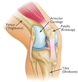
ANATOMY:
CLASSIFICATION:
SPECK and
REGAZZONI:
AO
Classification:
PATIENT INFORMATION:
Ø Patient
- ‘X’
Ø Age
- 75 years/ Female
Ø Date
of Admission: 05/12/2018
Ø Date
of Surgery: 07/12/2018
CHIEF COMPLAINT:
The patient had pain, swelling, and tenderness over the left knee (knee cap). The patient had restricted left knee movements
and inability to stand and walk few steps since fall (05/12/2018).
HISTORY
OF PRESENT ILLNESS:
The patient had restricted knee
movements and inability to use Left knee since fall. The patient gave a history of
slip and fall at the market. Since then
patient complaints of pain, swelling, range of movements restricted.
After the injury patient brought by
ambulance to the hospital. In the hospital,
the patient has been taken to the
emergency department. The patient was examined
by traumatologist and got admitted in the traumatology
department. X-ray was taken and it showed
transverse fracture patella with displacement.
v Previous
Injury: The patient gave a history
of fractured left wrist in 2017 treated
conservatively
v Developmental
History: No any developmental histories
v Drug
History: No known drug allergies. The patient is on treatment
for CVS.
v Past
Medical History: The patient gave a past medical history of Ischemic heart
diseases, Cardiosclerosis, CHF IIA, Arterial Hypertension II, Risk IV.
No DM; No Asthma; No thyroid disease.
v Past
Surgical History: No & No any blood transfusion.
ON EXAMINATION:
Patient is conscious, oriented.
Vital Signs:
· BP
– 130/80 mmHg
· PR
– 80/min
· SPO2
– 98%
LOCAL EXAMINATION:
· Pain
and Swelling over the Left knee are present.
· Tenderness
and Crepitus over the Left knee are present.
· The range of Motion of Left knee is restricted.
· Any
attempted movements painful.
· Active
toe movements present.
· Distal
pulse present.
X-RAY FINDING:
Transverse
Fracture Patella with displacement Left knee.
AP view:
Lat view:
PREPARATION:
Patient in Supine position:
Place the patient on a radiolucent table. A small bolster can be beneath the
buttock to prevent external rotation of the lower extremity.
SKIN INCISION:
Midaxial longitudinal
approach: Make a longitudinal mid-axial
incision from 5cm above the superior pole of the patella to the tibial tubercle.
PROCEDURE:
K-wire insertion :
Using the outside-in technique, drill the first
K-wire in an axial direction. The second K-wire is then drilled parallel to the
first, through the reduced fragments. It may be difficult to find the right
direction and position for the wires.
Two parallel K-wires should be inserted to give
more stable fixation.
Tension Band insertion:
POSTOPERATIVE
X-RAY:
Lat view:
AP view:
CLASSIFICATION:
Ø According
to AO Trauma Classification - C 1.1
Ø According
to Speck and Regazzoni Classification – B2
CONCLUSION:
Ø Early
surgery, good anatomical reduction, and
internal fixation help to recover the
full range of movements.
Ø Stability
is RESTORED.
--THE END--


Comments
Post a Comment
Thank you for your kind words and your support.