Trimalleolar CASE PRESENTATION
CASE PRESENTATION
INTRODUCTION:
Ankle fractures
are the most common injuries that are treated by most orthopedic surgeons. The
ankle is one of the strongest mortise joint (also known as the WOODWORK joints
or the talocrural joint). In the body, this is the formation of hinge joint by
the lower end of the tibia and the fibula that articulates with the talus.
Ankle fractures occur when one or both sides of the ankle are completely or
partially broken due to twisting injuries or fall injuries expressed during
play or sports.
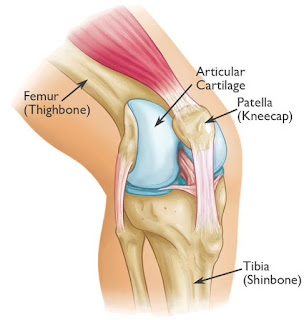
ANATOMY:

CLASSIFICATION:
Classification of ankle fractures is important
in order to estimate the extent of the ligamentous injury and the stability of
the joint.
The Weber classification focuses on the integrity of the syndesmosis, which holds the ankle mortise together.
The Lauge-Hansen system focuses on the trauma mechanism.
Adding the stages of Lauge-Hansen to the Weber system will help you to predict ligamentous injury and instability.
The Weber classification focuses on the integrity of the syndesmosis, which holds the ankle mortise together.
The Lauge-Hansen system focuses on the trauma mechanism.
Adding the stages of Lauge-Hansen to the Weber system will help you to predict ligamentous injury and instability.
Ø Weber A (Infrasyndesmotic)
– Lauge Hansen Supination adduction.
Ø Weber B (Transyndesmotic)
– Lauge Hansen Supination exorotation.
Ø Weber C (Suprasyndesmotic)
– Lauge Hansen Pronation exorotation.

PATIENT INFORMATION:
Ø Patient Name: “ X”
Ø Age: 48yrs/Female.
Ø Date of Admission:
05/04/2017.
Ø Date of Surgery:
08/04/2017.
CHIEF COMPLAINT:
Patient has
restricted right ankle movements and inability to stand and walk few steps
since today (05/04/2017).
HISTORY OF PRESENT ILLNESS:
Patient came with complaints of pain
,swelling over the right ankle and was unable to stand and walk few steps since
today (05/04/2017). Patient gave history of slip and fall at bathroom (at home)
today evening. Since then patient complaints of pain, swelling, deformity and
inability to use right leg (ankle). After the injury patient came to emergency
department of our hospital.
v Previous Injury: No
v Developmental History: No any developmental
histories
v Drug History: No known drug allergies.
Not on any chronic medication
v Past medical History: No Diabetes Mellitus; No
Hypertension; No Bronchial Asthma; No Thyroid
diseases.
ON EXAMINATION:
Patient is conscious,
oriented.
Vital
Signs:
·
BP – 140/90 mm/Hg
·
PR – 100b/min
·
SpO2 – 98%
·
Temp – 98° F
LOCAL EXAMINATION:
Ø Pain and swelling over the
right ankle (medial & lateral) is present.
Ø Tenderness and crepitus
over the right ankle (medial & lateral) is present.
Ø Deformity and angulation
over the right ankle (medial & lateral) is present.
Ø Any attempted movements of
the ankle are painful.
Ø Distal pulse palpable (A.Dorsalis Pedis).


X-RAY FINDING:
Before Reduction by
POP (Below Knee Slab)


After Reduction by
POP (Below Knee Slab)


Findings:
TRIMALLEOLAR
FRACTURE
1.
FRACTURE
DISTAL THIRD OF FIBULA RIGHT SIDE.
2.
FRACTURE
POSTERO MEDIAL ASPECT OF MEDIAL
MALLEOLUS RIGHT ANKLE
WITH SUBLUXATION OF
RIGHT ANKLE.
3.
UNDISPLACED FRACTURE BASE OF 2ND
,3RD & 4TH
METATARSAL RIGHT FOOT.
PREPARATION:
Supine Position by
Lateral and Medial Access:
The patient is
positioned supine on a radiolucent table with a sand bag under the ipsilateral
buttock. The injured leg may be placed over a folded blanket with the knee
slightly flexed. This position allows free access to both lateral and the
medial sides by hip rotation. Moving the leg towards the edge of the table
stabilizes the position of leg and ankle.

SKIN INCISION:
LATERAL INCISION:
The longitudinal lateral incision is the standard approach
for most lateral fractures.
*[If access to the anterior syndesmosis is
required, or a lag screw from anterior to posterior (Chaput lesion) is planned,
place the incision slightly anteriorly.]*
Make a 10-15 cm incision in line with the
fibula, centered over the fracture. If necessary, the incision may be extended
distally to reduce and hold the syndesmosis.

The fracture
site was exposed. Fracture was reduced.
Fracture was fixed with 7 holed reconstruction plate with 3 screws
proximally & 3 screws distally.
MEDIAL INCISION:
The incision started 2 cm distal
to the anterior tip of the medial malleolus. Curve the incision towards the
anterior edge of the medial malleolus and in the direction of the middle of the
distal tibia.

The fracture in the postero-medial
aspect was exposed. Fracture was reduced and was fixed with K-Wire. K-wire was
replaced with cannulated screws with washer.
The fracture reduction with
plate & screws checked under C-Arm & was found satisfactory.
AP view:

LAT view:

CLASSIFICATION:
According to the classification. The
fracture is classified:
Weber C This is a fracture above the level
of the syndesmosis. Usually there is a total rupture of the syndesmosis with
instability of the ankle.
According to Lauge-Hansen, it is the result of an exorotation force on the
pronated foot.

Ø Stage 1 - Avulsion of the medial
malleolus
or - ligamentous rupture
or - ligamentous rupture
Ø Stage 2 - Rupture of the anterior
syndesmosis
Ø Stage 3 - Fibula fracture above the level
of the syndesmosis (this is the true Weber C fracture)
Ø Stage 4 - Avulsion of the posterior
malleolus
or - rupture of the posterior syndesmosis
or - rupture of the posterior syndesmosis
POST-OPERATIVE
Range Of Movements:
14 Months after
Surgery:


DORSI FLEXION:

PLANTAR FLEXION:

CONCLUSION:
Ø Early surgery, good anatomical
reduction and internal fixation help to recover the full range of movements.
Ø Stability has been RESTORED.
--THE END--


Comments
Post a Comment
Thank you for your kind words and your support.