HIP DISLOCATION (FEMUR DISLOCATION)
HIP DISLOCATION (FEMUR
DISLOCATION)
INTRODUCTION:
A traumatic hip dislocation occurs when the head
of the thighbone (femur) is forced out of its socket in the hip bone (pelvis).
It typically takes a major force to dislocate the hip. Car collisions and falls
from significant heights are common causes and, as a result, other injuries
like broken bones often occur with the dislocation. A hip dislocation is a
serious medical emergency. Immediate treatment is necessary.
·
Epidemiology
Ø rare, but high
incidence of associated injuries
Ø mechanism is usually young patients
with high energy trauma
·
Hip joint
inherently stable due to
Ø bony anatomy
Ø soft tissue constraints
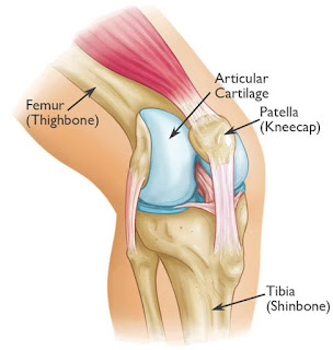
ANATOMY:

CLASSIFICATION:
Ø Simple:
dislocation without associated fracture.
Ø Complex:
dislocation associated with fracture of acetabulum or proximal fever.
v Anatomic
Classification:
Ø Posterior
Dislocation: (90%)

Ø
Anterior
Dislocation: (Rare)

SIGNS AND SYMPTOMS:
·
Acute
pain/ severe pain.
·
Inability
to walk and make few steps.
·
Deformity
·
If
there is any nerve damage (SCIATIC NERVE).
Patient will
have:
o
Numbness
over the foot and ankle.
o
Foot
drop.
o
Unable
to do dorsi-flexion of foot.
DIAGNOSIS:
Ø
X-ray:

Ø
CT-Scan:

Ø
MRI:
o
Useful to
evaluate labrum, cartilage and femoral head vascularity.
TREATMENT:
Ø NON-OPERATIVE:
o
Closed
Reduction: Reduction should be done within 6-8 hours.


Ø OPERATIVE:
o
Open Reduction
with or without removal of incarcerated fragments.
o
Open Reduction
Internal Fixation.
COMPLICATION:
Ø Osteonecrosis/Avascular Necrosis. (5-40%)
Ø Sciatic Nerve injury. (8-20%)
Ø Post-traumatic Arthritis. (Up to 20%)
Ø Recurrent Dislocation (Less than 2%)
--THE END--


Comments
Post a Comment
Thank you for your kind words and your support.