PILON FRACTURE
PILON FRACTURE
INTRODUCTION:
A pilon fracture is a type of break
that occurs at the bottom of the tibia (shinbone) and involves the
weight-bearing surface of the ankle joint. With this type of injury, the other
bone in the lower leg, the fibula, is frequently broken as well. A pilon
fracture typically occurs as the result of a high-energy event, such as a car
collision or fall from height.
Pilon is the French word for
"pestle"—an instrument used for crushing or pounding. In many pilon
fractures, the bone may be crushed or split into several pieces due to the
high-energy impact that caused the injury.
In most cases, surgery is needed to restore the damaged bone
to its normal position. Because of the energy required to cause a pilon
fracture, patients may have other injuries that require treatment as well.
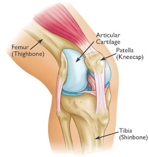
ANATOMY:
The two bones of the lower leg are the:
- Tibia—shinbone
- Fibula—smaller
bone in the lower leg
The talus is a small foot bone that works as a hinge between the
tibia and fibula. Together, these three bones—tibia, fibula, and talus—make up
the ankle joint.

DESCRIPTION:
Pilon fractures vary. The tibia may break in one place or
shatter into multiple pieces.
The severity of the injury depends on several factors, including:
- The
number of fractures
- The
amount and size of the broken bone fragments
- The
amount each piece is out of place (displaced)—In some cases, the broken
ends of bones line up almost correctly; in more severe fractures, there
may be a large gap between the broken pieces, or the fragments may overlap
each other.
- The
injury to the surrounding soft tissues, such as muscle, tendons, and skin
If the bone breaks in such a way that bone fragments stick out
through the skin or a wound penetrates down to the broken bone, the fracture is
called an "open" or compound fracture. This type of fracture is
particularly serious because, once the skin is broken, infection can occur in
both the wound and the bone. Urgent treatment is required to prevent infection.

CAUSES:
Pilon fractures most often result from high-energy trauma
such as a car or motorcycle accident, fall from height, or skiing accident.
Doctors have seen an increase in pilon fractures since the
introduction of air bags in motor vehicles. While air bags enable more people
to survive high-speed car crashes, they do not protect the legs—so many of the
survivors wind up with pilon fractures and other leg injuries.
SYMPTOMS:
Patients with pilon
fractures usually experience immediate and severe pain. Other symptoms may
include:
- Swelling
- Bruising
- Tenderness
- Inability
to bear weight on the injured leg
- Deformity—your
ankle may look angled or crooked
DIAGNOSTIC METHODS:
·
X-rays. X-rays provide images of dense structures, such as bone.
X-rays of the leg, ankle, and foot are commonly done to evaluate a pilon
fracture. An x-ray can show if there is an injury to your bones or if the
joints in your ankle are out of place.
·
Computed tomography (CT) scans. A CT scan can
provide valuable information about the severity of the fracture by helping your
doctor see the fracture lines more clearly. A CT scan will also help your
doctor plan your treatment. Your doctor may order a CT scan right away, or may
wait until later in your treatment—after an external fixator is applied.

TREATMENT:
·
Nonsurgical
Treatment
Nonsurgical treatment may be
recommended for stable fractures in which the pieces of bone are not displaced
or are minimally displaced.
It may also be recommended for patients who do not
do a lot of walking or for patients who are at higher risk for surgical
complications. For example, patients with severe osteoporosis, heart disease,
or other medical concerns may not be able to tolerate surgery.
Nonsurgical treatment may include:
Ø Splints
and casts. In most
cases, your doctor will first apply a splint to hold your ankle in place. Once
the swelling goes down, he or she will replace the splint with a short leg
cast. To provide effective support, your cast must correctly fit your ankle.
For this reason, as the swelling in your ankle decreases, you may need frequent
cast changes.
Ø Monitoring. Doctor will closely monitor the
healing of your fracture. During this time, you will need to return regularly
for follow-up x-rays to make sure your ankle remains stable.
Ø Recovery. You will most likely be unable to bear
weight on your ankle for up to 12 weeks after your injury. During this time,
your doctor may recommend that you use crutches or a walker. After 6 weeks,
your doctor may replace your cast with a removable brace. This will offer protection
while your ankle continues to heal
·
Surgical
Treatment
Surgery is commonly recommended for
unstable fractures in which the bones are out of place.
·
Open Reduction and Internal Fixation
During this operation, the displaced
bone fragments are first repositioned (reduced) into their normal alignment,
and then held together with screws and metal plates attached to the outer
surface of the bone.

·
Timing of Surgery
If you have significant swelling or
blisters, your doctor will delay your surgery until the swelling goes down.
Performing surgery too soon increases your risk for infection or problems with
your incision. Your surgery may be delayed for up to 2 weeks or more, depending
on how long it takes for the swelling to go down.
Doctor may place your ankle in a splint until
your surgery, or recommend that you have an initial smaller surgery to protect
your ankle while waiting for the second surgery.
·
External Fixation
Doctor may apply an external
fixator to hold your pilon fracture in place and stabilize your ankle until
your second surgery can take place.

REHABLITATION:
·
Weight
Bearing
Most patients are not able to put
all of their weight on their injured ankle for 2 to 3 months. Doctor may
recommend that you use crutches, a cane, or a walker during this time.
·
Physical
Therapy
Once you are allowed to start moving
your ankle, doctor may place it in a removable cast or brace so that you
can begin physical therapy. Specific exercises will help improve the range of
motion in your ankle. Exercises to strengthen the supporting muscles will be
added around 6 weeks after surgery.
As you transition to wearing regular shoes, you
will gradually stop wearing your brace. By 4 months after surgery, most
patients no longer need a walking aid.
Because your ankle can continue to improve for up
to 2 years, it is important to continue the exercises even after you have
completed the formal physical therapy program.
--THE END--


Comments
Post a Comment
Thank you for your kind words and your support.