GALEAZZI FRACTURE
GALEAZZI FRACTURE
INTRODUCTION:
1)
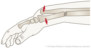
Definition
a)
distal 1/3 radius shaft FRACTURE AND
b)
associated distal radio-ulnar joint (DRUJ)
injury
2)
Incidence of DRUJ instability
a)
if radial fracture is <7.5 cm from
articular surface
i)
unstable in 55%
b)
if radial fracture is >7.5 cm from
articular surface
i)
unstable in 6%
3)
Mechanism
a)
direct wrist trauma
i)
typically dorsolateral aspect
b)
fall onto outstretched hand with forearm in
pronation
CLINICAL PRESENTATION:
1)
Symptoms
a)
pain, swelling, deformity
2)
Physical exam
a)
point tenderness over fracture site
b)
ROM
i)
test forearm supination and pronation for instability
c)
DRUJ stress
i)
causes wrist or midline forearm pain
DIAGNOSTIC METHODS:
1)
Radiographs
2)
recommended views
a)
AP and lateral views of forearm,
elbow, and wrist
3)
findings
a)
signs of DRUJ injury
i)
ulnar styloid fracture
ii)
widening of joint on AP view
iii)
dorsal or volar displacement
on lateral view
iv)
radial shortening (≥5mm)
TREATMENT:
1)
ORIF of radius
a)
approach
i)
volar (Henry) approach to radius
b)
plate fixation
i)
perform anatomic plate fixation of
radial shaft
ii)
radial bow must be restored/maintained

2)
Reduction & stabilization of DRUJ
a)
approach
i)
dorsal capsulotomy
b)
reduction technique
i)
immobilization in supination (6 weeks)
(1)
indicated if DRUJ stable following ORIF of
radius
ii)
percutaneous pin fixation
(1)
indicated if DRUJ reducible but unstable
following ORIF of radius
(2)
cross-pin ulna to radius
(a)
leave pins in place for 4-6 weeks
iii)
open surgical reduction
(1)
indicated if reduction is blocked
(a)
suspect interposition of ECU tendon
iv)
open reduction internal fixation
(1)
indicated if a large ulnar styloid fragment
exists
(2)
fix styloid and immobilize in supination


Comments
Post a Comment
Thank you for your kind words and your support.