Posts

Showing posts from September, 2017

AVASCULAR NECROSIS(AVN) OF THE HIP

Image
AVASCULAR NECROSIS (AVN) OF THE HIP INTRODUCTION:               Avascular Necrosis is otherwise called as Osteonecrosis or Aseptic Necrosis of the hip. AVN of the hip is a painful condition that occurs when the blood supply to the bone is disrupted. Because bone cells die without a blood supply, AVN can ultimately lead to destruction of the hip joint and arthritis.  Although it can occur in any bone AVN most often affects the hip. Many people each year enter hospitals for the treatment of AVN of the hip. In many cases, both the hips are affected by the disease. ANATOMY:             The hip is a ball-and-socket joint. The socket is formed by the acetabulum, which is part of the large pelvis bone. The ball is the femoral head, which is the upper end of the femur (thighbone). A slippery tissue called articular cartilage covers the surface of the ball...

DUPUYTREN’S CONTRACTURE

Image
DUPUYTREN’S CONTRACTURE INTRODUCTION:                     Dupuytren’s contracture is an abnormal thickening of the tissue just beneath the skin. This thickening occurs in the palm and can extend into the fingers. Firm pits, bumps and cords (thick lines) can develop and cause the fingers to bend into the palm.This condition may also be known as Dupuytren’s Disease. Occasionally, the disease will cause thickening on top of the knuckles or cause lumps and cords on the soles of the feet (plantar fibromatosis). ANATOMY:   SYMPTOMS:       A Dupuytren's contracture typically progresses very slowly, over a period of years. Signs and symptoms of the condition may include: Ø   Nodules:   You may develop one or more small lumps, or nodules, in the palm of your hand. These nodules are typically fixed to the overlying skin. Initially, the ...

Avulsion Fracture PCL Injury

Image
Avulsion Fracture  Posterior Cruciate Ligament Injury The posterior cruciate ligament is located in the back of the knee. It is one of several ligaments that connect the femur (thighbone) to the tibia (shinbone). The posterior cruciate ligament keeps the tibia from moving backwards too far. An injury to the posterior cruciate ligament requires a powerful force. A common cause of injury is a bent knee hitting a dashboard in a car accident or a football player falling on a knee that is bent. Anatomy Two bones meet to form your knee joint: your thighbone (femur) and shinbone (tibia). Your kneecap sits in front of the joint to provide some protection. Bones are connected to other bones by ligaments. There are four primary ligaments in your knee. They act like strong ropes to hold the bones together and keep your knee stable.    Normal knee anatomy, front view :         Collateral ligaments.  These are found on the ...

SMITH’s FRACTURE

Image
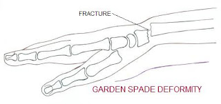
SMITH’s FRACTURE INTRODUCTION:                             Smith’s Fractures is also known as a Goyrand Fracture in French literature. Smith’s fracture is palmarly displaced distal radius fracture. It is referred to as “ GARDEN SPADE ” deformity. Smith’s fracture is a reversed Colle's fracture.      ANATOMY: EPIDEMIOLOGY :                            Smith’s fracture account for less than 3% of all fractures of the radius and ulna and have a bi-modal distribution: young males (most common) and elderly females. MECHANISM: Smith’s fractures usually occur in one of the 2 ways: Ø   a fall onto a flexed wrist Ø   direct blow to the back of the wrist ...